What Is a Stroke Radiology Assistant?
A stroke radiology assistant is a trained professional who supports imaging teams by preparing patients, assisting during brain scans, and helping doctors quickly identify signs of a stroke. For a clearer understanding of responsibilities and training, please go through the details below.
When every heartbeat matters during a medical crisis, fast and accurate diagnosis can decide whether someone regains full strength or faces lasting difficulties. Behind urgent moments in emergency wards or neurological units stands a focused expert, ensuring vital images are captured exactly when needed.
While clinicians handle direct treatment, these imaging specialists work in silence, keeping each scan sharp, quick, and precise. Their mastery of advanced equipment turns complex technology into a lifeline during chaos. Exploring their work reveals how steady precision and timing can steer critical outcomes when every second carries weight.
What Is a Stroke Radiology Assistant?
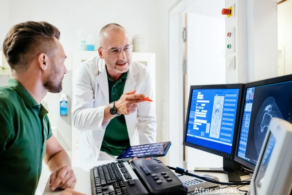
They’re highly trained professionals who work closely with doctors, using advanced imaging tools like CT, MRI, or angiography machines. Their job includes getting patients ready, capturing clear scans, organizing image files, and occasionally offering a first look before a full review is done.
Unlike others in similar roles, they’re well-versed in brain anatomy, warning signs, and key imaging details tied to blood flow issues in the brain. Their specialized experience helps them work quicker and with greater precision—something that’s absolutely crucial when every second counts.
Role in Stroke Diagnosis
When it comes to diagnosing brain emergencies, imaging plays a crucial role. There are typically two main types of issues in these situations:
One happens when a clot blocks normal flow toward brain tissue, stopping delivery of oxygen and nutrients it depends on. As circulation slows or shuts down, affected areas begin to struggle, and cells lose ability to function properly. Without timely restoration of flow, damage can spread beyond initial spot, leading to lasting effects tied to how long interruption continues.
Another happens when a vessel breaks open, allowing blood to spill into or around brain regions. Pressure then builds inside enclosed space, interfering with normal activity nearby. As fluid spreads, surrounding areas become irritated or compressed, which can quickly worsen effects if action does not follow soon.
These two conditions show up very differently on scans, which makes quick identification absolutely essential.
Imaging Techniques Used in Stroke Care
Those who specialize in brain-related emergencies need to become highly skilled with several imaging methods:
CT Scans (Computed Tomography)
This approach usually comes first and delivers results quickly when looking for bleeding inside brain areas. It allows rapid visualization, which matters when time feels critical. Their role focuses on careful positioning of a person, making sure movement stays minimal throughout scanning. Attention also goes toward choosing proper scan settings, since accuracy depends on correct angles, timing, and clarity. Through steady coordination and technical precision, they ensure images come out sharp and reliable, giving professionals dependable information to guide next decisions.
MRI (Magnetic Resonance Imaging)
This technique delivers clearer, more refined views of brain structures, making early signs of blockages easier to notice. It demands strong expertise to decide which scan styles fit best, such as diffusion-weighted imaging, since each option highlights changes in a different way. Through informed selection and precise execution, it becomes possible to reveal subtle problem zones that might otherwise remain hidden, allowing findings to emerge with greater clarity and confidence.
Angiography (CT or MR Angio)
This approach maps blood vessels, allowing identification of clots or weak spots along circulation paths. Responsibilities involve handling contrast dye with precision, timing scans accurately, and fine-tuning settings so images appear sharp and easy to interpret. Careful coordination across each step ensures results reflect vessel structure clearly and reliably.
Perfusion Imaging
This examines how blood moves through brain tissue over time. It requires exact timing, careful contrast handling, and strong familiarity with specialized software. They receive thorough training for this work, allowing smooth coordination of each step and accurate interpretation of flow patterns as results appear.
Training & Education
Entering this profession often starts with certification as a general technologist through ARRT, American Registry of Radiologic Technologists. After qualification, individuals build experience on site and move toward advanced positions or focused credentials that match personal interests and strengths.
Education paths usually cover several core areas, allowing learners to understand how complex systems function and respond during critical moments. Coursework often explores structure and function related to brain and nerve activity, along with explanation of how various conditions develop and influence daily function. Instruction also emphasizes safety practices, proper use of contrast materials, and rapid decision-making during urgent imaging situations. Training frequently includes guidance on operating within specialized units, focusing on coordination, accuracy, and efficient task flow.
Many medical centers also offer customized in-house instruction designed specifically for this specialty. Such programs concentrate on practical challenges encountered during real scenarios and refine skills required for each position. Through continued learning and hands-on exposure, individuals steadily grow confidence and readiness for higher responsibility roles.
Collaboration with Healthcare Teams
Managing such conditions depends on tightly coordinated teamwork across multiple roles. Everyone involved must exchange information rapidly and with precision, since timing influences every decision made during urgent moments. Clear updates move between emergency room physicians so imaging findings reach decision-makers without delay. Neurology specialists rely on immediate communication to recognize brain-related patterns and guide next steps. Radiology professionals coordinate closely to prepare scans, set priorities, and keep workflow moving smoothly. Nursing teams within intensive units and neuro wards align transport timing and maintain safety during movement. Emergency medical service teams stay informed so urgent protocol activation happens without confusion.
Across many facilities, this specialized group becomes active during high-priority alert activations. Such situations unfold at a rapid pace and leave little room for hesitation. Everyone involved depends on quick diagnostic action, precise coordination, and steady communication to keep each step aligned from arrival through imaging and onward decision points.
Stroke Types & Radiological Indicators
They do not interpret scans or deliver formal conclusions, yet recognition of critical visual cues remains essential. Spotting specific patterns allows quick adjustment of protocols and signals when immediate communication with other professionals becomes necessary. Awareness at this level improves speed, coordination, and overall efficiency during urgent situations.
Several indicators commonly appear during imaging review. One example involves a hyperdense MCA sign, which often suggests early ischemic changes. Another involves midline shift, usually linked with swelling or internal bleeding that pushes structures away from normal alignment. Effacement of sulci can signal increased swelling that compresses natural grooves along brain surfaces. Contrast leakage may appear when bleeding allows injected material to escape expected pathways. Loss of gray-white differentiation often reflects early ischemic change, where normal visual separation between tissues fades.
Understanding how these signs appear—without assigning a diagnosis—allows personnel to act decisively. This visual familiarity helps them escalate concerns quickly, streamline workflow, and communicate urgency with clarity during time-sensitive scenarios.
Speed & Accuracy in Emergency Settings
Each passing minute of delay allows nearly two million neurons to perish, which explains why speed paired with precision remains non-negotiable. Timing influences outcomes directly, so actions must stay focused and efficient from first contact onward.
Individuals serving in this position receive training that emphasizes rapid execution without sacrificing safety. Emphasis remains on limiting unnecessary movement, especially when working with older adults who may experience fragility or instability. Quick yet secure positioning becomes essential, allowing imaging to begin without hesitation. Close coordination with emergency room teams ensures imaging takes priority during urgent arrivals. Preparation of contrast agents happens within moments, requiring familiarity, confidence, and steady hands. Readiness also extends to unexpected emergencies, including allergic responses or sudden code situations, where immediate reaction can make a critical difference.
Across certain facilities, protocols require completion of CT imaging within twenty-five minutes after arrival. Meeting such benchmarks depends heavily on professionals in this role, since efficiency, preparation, and communication directly influence whether that target gets achieved.
Technology & Tools in Radiology
Individuals in this position must remain highly tech-savvy, since daily tasks involve constant interaction with advanced digital tools. Work often includes navigation of PACS platforms, where images get stored, retrieved, and shared within seconds. Imaging software such as RAPID or Viz.ai assists with AI-enhanced detection, requiring familiarity with alerts, overlays, and automated flags. Operation of CT and MRI control panels demands precision, speed, and confidence, especially during urgent scans. Use of contrast injectors requires accurate setup, timing, and monitoring. Digital records and EMR platforms also play a major role, since documentation must stay accurate and immediately accessible.
Alongside equipment handling, continuous observation of individuals undergoing scans remains essential. Monitoring becomes especially important when sedatives or IV contrast agents come into play. Attention stays focused on breathing patterns, responsiveness, and any sudden changes that may signal distress. This combination of technical proficiency and real-time observation ensures procedures move forward smoothly while maintaining safety throughout imaging sessions.
Case Studies & Real-Life Examples
Case 1: Middle-of-Night Emergency
An elderly man arrives around 2 AM showing slurred speech and sudden confusion. She moves fast, escorts him straight to imaging, sets up equipment, and completes scanning in under ten minutes. Her preparation stays sharp despite late hour pressure. Images quickly reveal a blockage within a major artery. Thanks to her speed and precision, medication gets delivered within a critical time window. Swift action changes outcome, proving how accuracy during urgent moments saves irreplaceable function.
Case 2: Child in Trouble
A young boy arrives displaying confusion and noticeable weakness. He steps in to assist during a sedated scan, remaining calm and attentive throughout process. Clear images uncover a rare brain condition requiring immediate attention. His steady voice and reassuring manner ease fear for both child and anxious parent. Because of this composed approach, procedure continues smoothly and safely, allowing clinicians to move forward with confidence and clarity.
Why Stroke Radiology Assistants Matter
Most people rarely notice them, yet their work underpins every life-saving moment. From preparing machines to capturing images relied upon by doctors, they play an essential role.
During moments when each second carries weight, their actions lead to meaningful improvements in outcomes. Quick thinking guides decisions, refined skills keep tasks precise, calm focus holds steady under pressure, and close attention prevents missteps. Working away from spotlight, they operate as a quiet force, shaping real change through timing, accuracy, and unwavering dedication in situations where every choice truly matters.
Final Thoughts
When someone survives a major medical emergency thanks to rapid action and precise diagnosis, consider professionals working behind scenes to prepare critical scans. Their work may go unnoticed, yet impact remains vital.
For anyone drawn to fast-moving, life-saving settings without a need for spotlight attention, this profession offers a powerful and rewarding path. Work happens behind scenes, yet impact remains immediate and profound. In this role, each second holds weight, and every image captured carries real meaning—shaping decisions, guiding action, and influencing outcomes when timing matters most.
Frequently Asked Questions About Diagnostic Imaging Professionals and Their Role in Patient Care
How do professionals in diagnostic imaging prepare patients for scans?
Professionals ensure patients sit or lie correctly and remain comfortable before any scan. They walk through each step using clear, simple language to ease nerves, especially for people unfamiliar with equipment or techniques. Preparation also involves actions such as adding contrast material when required or removing metal items to prevent interference. This thoughtful setup plays a vital role in producing sharp images while keeping everyone safe during process.
What role does technology play in improving patient care during imaging?
Modern diagnostic equipment is highly advanced, using software that sharpens image clarity and precision. Professionals depend on these tools to capture high-quality scans rapidly, some with automated systems analyzing images in real time. Such innovations enable faster diagnosis and support critical decision-making during emergencies. They also collaborate with AI-powered technologies that highlight conditions not immediately obvious, improving overall accuracy and efficiency of each scan.
How do professionals handle emergencies or urgent cases?
In urgent cases, speed is crucial. When a patient shows signs of a neurological problem, imaging professionals act swiftly to capture required scans. They follow protocols to prioritize critical situations, ensuring procedures are carried out efficiently. Coordination with emergency staff or neurologists ensures results reach decision-makers immediately, enabling rapid, informed action. Expertise in staying calm and maintaining organized workflows is essential in high-pressure environments.
What are some common challenges faced during imaging procedures?
Even with advanced imaging technology, challenges remain. Some patients struggle to stay still due to pain, anxiety, or medical conditions, which can create blurry images and necessitate repeat scans. Claustrophobia, especially during MRI procedures, may require extra attention or sedation. Timing contrast injections accurately and preparing patients properly for specific imaging types adds further complexity. Professionals work diligently to overcome these obstacles, ensuring imaging quality remains high and reliable.
How do professionals ensure the safety of patients during imaging?
Patient safety stays a top priority. Professionals review each patient’s medical history for potential risks, including allergies to contrast agents. They monitor for adverse reactions during procedures and apply proper safety measures. Radiation exposure is minimized by using lowest effective doses while strictly following safety protocols.
What is the role of professionals in coordinating with other healthcare providers?
Professionals specializing in diagnostic imaging collaborate closely with doctors, neurologists, and surgeons. They share scan results and offer insights into findings, aiding in more informed treatment decisions. Clear communication with medical teams ensures procedures proceed efficiently, supporting smooth coordination of patient management.
How do professionals in imaging handle pediatric patients?
When working with pediatric patients, professionals take extra measures to make procedures as stress-free as possible. They often use child-friendly equipment designed for comfort and smaller size. Procedures are explained in simple, reassuring terms to help children feel at ease. Sedation may be used for younger or anxious patients to ensure they stay still during scans. Every step focuses on maintaining safety and comfort while capturing accurate images.
What training and certifications are required to work in this field?
Professionals working within diagnostic imaging often begin by earning certification in radiologic technology. This initial credential opens entry into practice and builds a strong technical foundation. After gaining experience, many choose to deepen expertise through specialization in specific techniques or focused areas, including neurological imaging or pediatric imaging, depending on interest and workplace needs.
Further credentials may become necessary based on equipment used or responsibilities assigned. Advanced scanners, specialized software, or leadership duties often require additional training and formal recognition. Learning does not stop after certification, since ongoing education plays a central role in keeping skills current. Imaging tools, digital systems, and procedural standards continue to evolve, and staying informed ensures accuracy and confidence during daily tasks.
These structured learning paths shape technical ability, decision-making speed, and professional growth. Through continued study and hands-on experience, individuals maintain proficiency and remain effective within a field defined by constant change and advancing innovation.